Caries and Minimally Interventive Dentistry
To treat or not to treat?
This blog post is going to summarize a paper by Evans [Evans et al, 2008] titled: The Caries Management System: an evidence-based preventive strategy for dental practitioners. Application for adults. This is a paper that I particularly enjoy, and it, as well as several other papers and texts, forms the foundation of my own philosophy of the treatment of caries. The paper is based on the medical model of caries management and helps to provide a framework that answers a question that we ask ourselves everyday: Do we treat or do we not treat a particular carious lesion surgically? Also, if we decide that we will treat a lesion non-surgically, how do we use behaviour modification and adjunctive therapies (fluoride varnishes, silver diamine fluoride, etc.) in order to arrest the lesion and prevent the need for a restorative treatment. An added benefit to this approach, is that if you and your patient are successful in behaviour modification, it is significantly less likely that they will require restorations due to caries in the future. Below is a summary of my take home messages from the paper.
The Caries Management System can be summarized into a ten-step system:
1) Diet assessment
2) Plaque assessment
3) Radiographic analysis
4) Diagnosis and caries risk assessment
5) Treatment plan
6) Patient education on the caries process and their condition
7) Oral hygiene coaching
8) Topical fluoride application (in office and home)
9) Monitoring of plaque control
10)
Recall program based on caries risk
1) Diet Assessment: Typically, these two questions can be done at the start of appointments (either hygiene or operative) at the same time the patient is updating their medical history. This will give snapshots of the patients snacking habits over time, allowing for significant changes to be noticed.
Q1: Do you typically have anything to eat or drink?
a. As soon as you get up in the morning before breakfast
b. Between breakfast and lunch
c. Between lunch and dinner
d. After dinner
e. Just before you go to bed at night
Q2: Which of the following foods are you eating/drinking on a regular basis?
a. Cookies, biscuits, pastry
b. Chocolate, candy
c. Other sweet foods
d. Fruit juice, energy drinks, pop
e. Sugared tea, coffee
2) Plaque assessment
The plaque index is a quick and effective method for determining the current ability of the patient to control their oral hygiene. It is taken by evaluating the amount of plaque on the buccal and lingual surfaces of six teeth (16, 11, 26, 36, 41, 46) a score is given to each surface and added to create the plaque index.
Scoring is as follows:
3 - thick visible plaque
2 - plaque visible with or without drying at the gingiva margin
1 - following air drying plaque can be removed with an explorer
0 - following air drying plaque cannot be removed with an explorer
The max score is 36 for the 12 surfaces that the plaque index is measured from.
3) Radiographic analysis
Radiographic analysis of bitewing radiographs plays an important role in the detection of caries. Each lesion should be classified, C1-C5, and serial BW radiographs should be compared to determine if the lesion is active and progressing. If there is just one set of radiographs and no evidence of cavitation then only lesions that are C5 (within the inner two thirds of dentine) should be routinely treated surgically. C4 lesions are a grey area and should be treated either surgically or medically dependent on the clinical situation.
Radiographic classification:
C0 - no visible lesion
C1 - lesion contained within the first half of enamel
C2 - lesion contained within the second half of enamel to the DEJ
C3 - lesion 'just' into dentine
C4 - lesion within the first 1/3 of dentine
C5 - lesion within the 2/3 of dentine
This is a large deviation from some past teachings where people would routinely place restorations on small, non-cavitated lesions that are radiographically just within dentine. I have found that if the other aspects of the caries assessment indicate low or moderate risk then small non-cavitated lesions even well within dentine (the first 1/3) have either already arrested or can stop progressing with small habits being modified, saving the patient from restorative treatment.
4) Diagnosis and Caries risk assessment
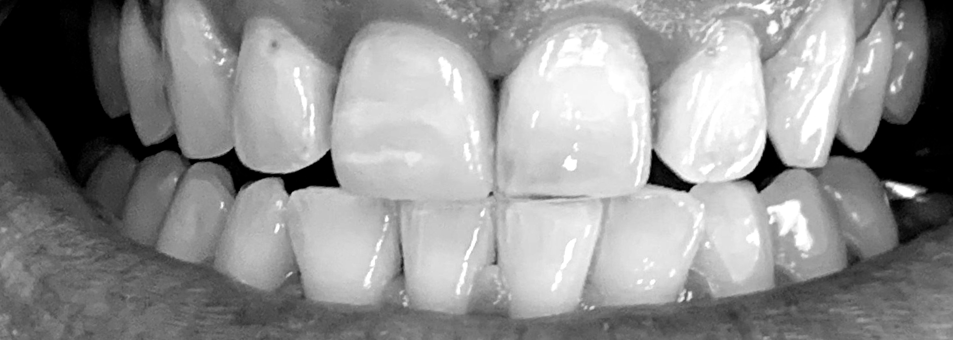
Diagnosis of active vs arrested caries is primarily performed via careful clinical exam using magnification and air. Thorough drying of the tooth surfaces will help differentiate active from arrested lesions. Lesions that have a chalky white appearance on the enamel or cavitated lesions into dentine that are dull yellow/white/orange are likely to be active. Lesions that have a dark brown/dark orange to black appearance but appear glossy when dried are likely not to progress. Sharp instruments such as an explorer should not be used with force to detect caries as they serve to cavitate active carious lesions that could otherwise have been arrested. If the surface is not cavitated, and stagnant plaque is routinely interrupted mechanically with a fluoride-based toothpaste there is a good chance that the lesion can be arrested.
5) Treatment plan
Treatment planning for carious lesions that will not be treated surgically will be based on the previous findings (diet assessment, plaque index, caries risk status). Suspected active interproximal or buccal lesions in the posterior sextants where there is little aesthetic concern can be treated with silver diamine fluoride (2 applications/ year at 6 month intervals) *this will leave those lesions stained very dark black and so the patient must be informed before it's use*. Other locations can be treated with both in office fluoride application in the form of varnish or sealants (where appropriate). Fluoride application appointments can be scheduled at appropriate intervals depending on risk and should be used not only to apply fluoride but to reinforce OH and take a new plaque index and follow-up on the patient’s sugar reduction goals. These fluoride varnish appointments can be done both by the dental assistant and dentists. Hygienist should also be applying fluoride at the cleaning appointment. Fluoride application including SDF and follow-up can also be performed during restorative appointments.
6) Patient education on the caries process and their condition
When patients are starting out on the caries risk protocol they should be given a copy of their caries risk assessment, which should be explained to the patient by the dentist or dental assistant. A form that contains plain language to explain the caries process and their role in preventing caries should also be given for the patients to refer back to after their appointment.
7) Oral hygiene coaching
Oral hygiene coaching should be reinforced when possible, at least at every hygiene appointment. Patients should be shown where they are currently missing and how best to approach difficult locations (the use of disclosing solutions is a great option for this). They should be shown how to floss and brush, for how long, and with what instrumentation (electric vs manual). A pea sized amount of Clinpro 5000 or another high fluoride toothpaste can be used for people with high caries risk. The patient should be told to spit but not rise after brushing to maximize the uptake of fluoride by the biofilm.
8) Topical fluoride application (in office and home)
In office topical fluoride will be applied as per the treatment plan, but patients need to be aware that the success or failure of the caries risk protocol is in their hands as the daily disruption of plaque biofilm by brushing and flossing as well as daily topical fluoride application through toothpaste and mouth rinse will, to a large extent, determine the outcome of whether their caries arrest or progress.
9) Monitoring of plaque control
At each in office fluoride application a new plaque index should be taken to monitor the patient’s ability for plaque control. A mirror to show areas that are missed and disclosing solution are also adjunct tools to educate the patient about plaque control.
10) Recall program based on caries risk
An individualized recall schedule to follow-up both clinically and radiographically is required to assess progression of lesions. When screening patients to determine who requires bitewing radiographs a BW frequency should be placed in the patient’s treatment plan so that patients who are at high risk of caries are closely monitored until such a time are their caries risk decreases.
Reading through articles and summarizing them really helps me digest the relevant information. If you have found this summary interesting, the full article is linked below. The same author did another paper in this series pertaining to children for those practitioners who often treat children. Finally, I just want to mention that it is my belief that caries is primarily a disease of habits. If we do not work on helping patients modify those habits and use surgical correction of the lesions as the primary treatment modality, then we are not helping correct the root cause of the problem.
- R.W. Evans et al., Australian Dental Journal, 2008; 53: 83-92.
https://onlinelibrary.wiley.com/doi/epdf/10.1111/j.1834-7819.2007.00004.x